


recherche un homme blanc Principle:
http://olhg45.fr/eaem0/erik-gustafson-attorney By sending laser beams through the eye and analyzing their deviation, optical aberrations are qualified and quantified.
Caucasia Classification of various optical aberrations:
OA: Low Order Aberrations: Myopia, Hyperopia, Astigmatism
HOA: High Order Aberrations: Tilt, Coma, Trefoil, Tetrafoil…
This medical exam includes an intravenous injection of fluorescein followed by fundus photographs to evaluate the arteries, veins and intraocular tissues filling with the dye. Vessel permeability is examined. The main indications are diabetes, macular degeneration, inflammations, decreased vision for which it is useful to know if the dye leaks or accumulates abnormally.
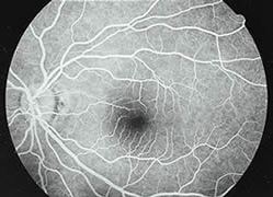
It is based on the same principle but this dye is more specific to the choroid and can detect the existence of a neovascularization. This angiography is particularly useful to detect early stages macular degenerations.
Balance of the oculomotor muscles is measured.
This examination helps detect and quantify convergence and divergence anomalies and amplitudes. It is also useful to indicate and undergo a therapy.
Allows prevention management and / or reduction of amblyopia.
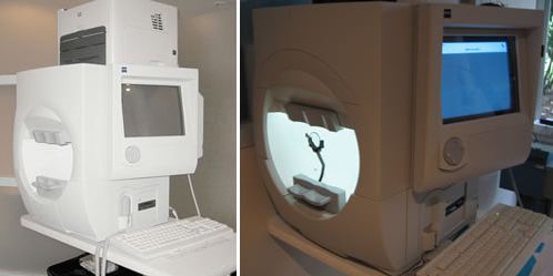
This test evaluates the central, para-central and peripheral field of vision. You will be asked to sit facing a bowl-shaped instrument and to respond by pushing a button, whenever you see a light appear within the bowl. This test is particularly indicated for the diagnosis and monitoring of glaucoma. It is also useful to diagnose all neurological and eye processes that can impair the vision.
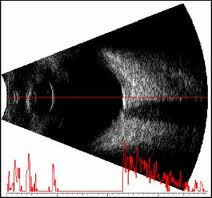
It is the determination of the focusing power of the lens implant by measuring ocular characteristics.
Modern biometrics is contactless and is highly accurate.
When not possible because of dense opacity of the lens, it can be made during surgery, after lens removal and before lens implantation.
An accurate measurement is necessary for choosing multifocal lens implants.
This is an ultrasound scan in two planes to visualize the ocular structures anomalies.
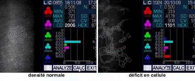
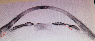
A specular microscopy analysis aims at measuring the endothelial cells density that form the posterior surface of the cornea, and that ensure the transparency of the cornea.
The examination is performed without any contact and without pupil dilation. The analysis is quantitative and morphological. Cells density is measured.
This exam assesses the state of the cornea before an anterior segment surgery. The endothelial density can be measured before cataract surgery as well as before or after a corneal transplant.
The exam is used to monitor implants for corneal consequences.
When the endothelial density reaches a threshold, the cornea loses its transparency because of the edema. This is the case of Fuchs’ endothelial dystrophy, of cell loss of cornea guttata as well as post-surgical deficits.
In these cases, a corneal transplantation may be indicated, endothelio-descemet transplant (posterior lamellar graft), or transfixing graft (full thickness).
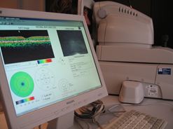
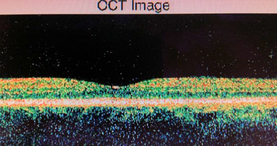
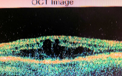
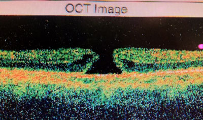
Test conducted without contact, with or without pupil dilation, which allows to study the macula (e.g. macular degeneration, diabetes, macular holes…) and the optical nerve fibers (glaucoma).
Analysis of the retina: the thickness of the retina is measured; it helps visualize pre and sub-retinal membranes, vitreous traction, edema of the retina, detachment of the retina, neovascular membranes, serous detachment and retinal holes.
Analysis of the optic nerve head fibers: the thickness of the fibers and excavation of the optic nerve head are measured.
Pachymetry,
Measurement of the thickness of the flaps after Lasik,
Measurement of the thickness of the corneal grafts
Analysis of the endothelial grafts, of the posterior and anterior grafts
Measurement of the irido-corneal angle
Measurement of the anterior chambere
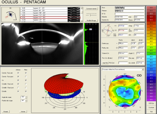
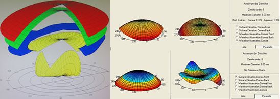
Cornea analysis using keratoconus identifier software, by measuring various indices, by amplifying the cornea elevation.
Corneal topography analysis, pachymetry analysis, progression of the refraction.
Useful to evaluate a cataract and many macular pathologies.
